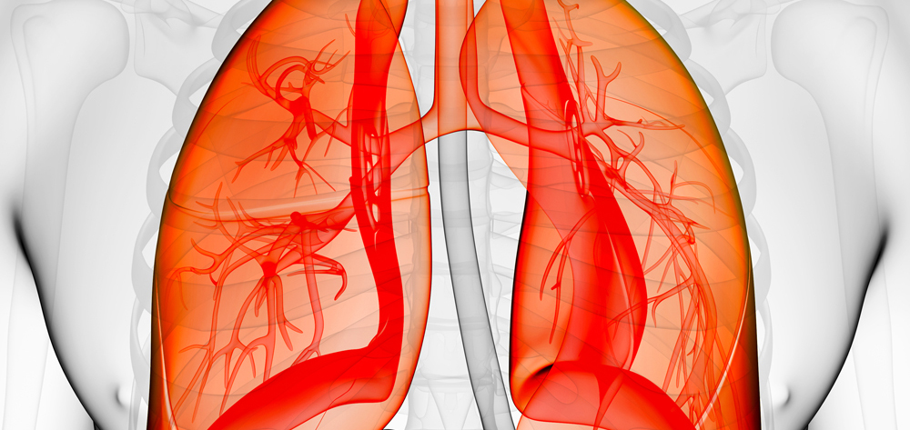
In the United States, patients with acute respiratory distress syndrome (ARDS) occupy 1 in 10 critical care beds. Each year, ARDS kills 150,000 Americans. Most of the deaths are triggered by an event, such as sepsis or pneumonia.
What is ARDS?
Usually, ARDS starts with acute lung injury (ALI), which kills about 75,000 Americans a year and whose risk increases with age. ARDS itself develops from an injury to the alveoli, the site of gas exchange. ALI and ARDS have these characteristics in common:
- Acute onset
- Bilateral infiltrates on chest X-ray
- Pulmonary artery wedge pressure (PAWP) of less than 18 mm Hg.
What distinguishes ALI from ARDS is the P/F ratio, the comparison of arterial partial pressure of oxygen (PaO2) with inspired fractional concentration of oxygen (FiO2). Simply put, the P/F ratio is a comparison of the amount of oxygen given to a patient with the amount of oxygen actually entering the patient’s bloodstream.
Understanding noninvasive ventilation
Evidence-based update on chest tube management
When breathing is a burden: How to help patients with COPD
The higher the P/F ratio, the better the gas exchange. The normal measurement is around 500 mm Hg. A P/F ratio below 300 mm Hg regardless of the positive end-expiratory pressure (PEEP) measurement indicates ALI. A P/F ratio below 200 mm Hg regardless of the PEEP measurement indicates ARDS.
If a patient’s oxygen requirements continue increasing while oxygen saturation levels (based on finger-probe readings and arterial blood gas [ABG] measurements) remain low, ALI is progressing to ARDS. This condition is called refractory hypoxemia.
A patient with ARDS has decreased functional residual lung capacity, which may lead to organ failure and death. Typically, ARDS requires admission to an intensive care unit (ICU) and mechanical ventilation.
What’s the damage?
ARDS is marked by inflammation, increased permeability of alveoli membranes, and cytokine activation. Damage to the endothelium, or lining of the capillary, interferes with alveolar gas exchange. The damage causes macrophages to release cytokines, inflicting more damage on the alveolar endothelium. Protein levels build, pulling fluid into the alveolar spaces and causing noncardiac pulmonary edema and a reduced surfactant level.
Normally, surfactant decreases surface tension and allows the alveoli to open easily. In ARDS, the edema and reduced surfactant level compromise gas exchange, causing decreased oxygen and increased carbon dioxide in the blood. The result is hypoxemia, pulmonary hypertension, and decreased pulmonary compliance. In later stages of ARDS, progressive alveolitis and fibrosis—stiff lungs—further decrease pulmonary function.
Signs and symptoms
In the first 24 to 48 hours, signs and symptoms include dyspnea, tachypnea, dry cough, fatigue, and tachycardia. Even with supplemental oxygen, a patient’s skin may look cyanotic and mottled. Auscultation reveals adventitious breath sounds (crackles, rhonchi, and wheezes) or increasingly diminished breath sounds. As oxygenation and perfusion diminish, the patient may become agitated, anxious, confused, and restless.
A chest X-ray shows diffuse infiltrates, and ABG results indicate respiratory alkalosis with very low PaO2 levels. In the later stages, hypercapnia may develop. Further metabolic imbalances can lead to mixed acidosis, signaling a low ventilation-to-perfusion (V./Q.) ratio and a deteriorating P/F ratio. To rule out a cardiogenic cause of pulmonary edema, a physician may order PAWP measurements.
5 P’s of ARDS therapy
Managing patients with ARDS requires maintaining the airway, providing adequate oxygenation, and supporting hemodynamic function.
The five P’s of supportive therapy include perfusion, positioning, protective lung ventilation, protocol weaning, and preventing complications.
Perfusion
The goal of care for ARDS patients is to maximize perfusion in the pulmonary capillary system by increasing oxygen transport between the alveoli and pulmonary capillaries. To achieve the goal, you need to increase fluid volume without overloading the patient. Give either crystalloids or colloids to replace the fluids that have leaked from the capillaries into the alveolar spaces. Blood transfusions can improve oxygen delivery, but remember they can also cause an increased inflammatory response and increase the risk of infection and death.
Evaluate the patient’s volume status by measuring blood pressure, respiratory variations of pulmonary and systemic arterial pulse pressure, central venous pressure, and urine output. Confirm intravascular status with pulmonary artery catheter data, cardiac output, cardiac index, pulmonary vascular resistance, and venous oxygen saturation (SvO2).
Certain drugs can also help increase perfusion. Inotropics such as dobutamine (Dobutrex) can increase cardiac output to boost oxygenation. Milrinone lactate (Primacor), another inotropic, improves perfusion by causing vasodilation in the pulmonary bed
Vasopressors, such as norepinephrine and dopamine, promote systemic vasoconstriction, thus increasing blood pressure and perfusion. When administering these drugs, monitor vital signs, skin color and temperature, and the patient’s tolerance to therapy.
Positioning
Patient positioning also affects perfusion. If a patient is standing, blood flow moves to the base of the lung and away from the apex. If a patient is supine, the posterior area of the lung will be more perfused than the anterior area. Because the better aerated surfaces of the lungs are the nondependent areas, the result is a ventilation/perfusion mismatch.
The same thing happens with PEEP, which primarily aerates the anterior, nondependent areas of the lungs instead of the dependent areas that would benefit most.
Immobility, a major cause of pulmonary complications, greatly influences perfusion distribution. Three positioning therapies can decrease these complications and improve perfusion in ARDS patients:
- Kinetic Therapy (bilateral turning of a patient 40 degrees or more per side)
- continuous lateral rotational therapy (bilateral turning of a patient no more than 40 degrees per side)
- prone positioning.
These therapies improve oxygenation by mobilizing secretions, resolving atelectasis, improving V./Q. ratio, recruiting functional but collapsed or consolidated alveolar units, and decreasing interstitial fluid accumulation. Rotational therapy reduces nosocomial pneumonia, skin breakdown, ICU length of stay, and the number of ventilator days.
There’s a correlation between the degree of rotation and the therapeutic benefit. With rotational therapy, the patient must be turned consistently to 40 degrees; otherwise, the therapy doesn’t significantly affect the clearance of secretions, risk of pneumonia, or the length of ICU stay. Kinetic Therapy is effective in immobilized patients at angles up to 62 degrees.
Kinetic Therapy effectively prevents and treats severe respiratory complications of prolonged immobilization. When started early, it prevents and treats pneumonia and ARDS, saving hospital resources and lives.
Using the prone position
Prone positioning improves the V./Q. ratio. Aeration improves because the heart no longer compresses the posterior areas of the left lung as it does in the supine position. With the patient in the prone position, most lung tissue, which is in the posterior areas, moves toward the anterior, clearing the airways of debris, decreasing atelectasis, reducing lung inflammation, and producing more efficient oxygenation and perfusion.
Prone positioning can be achieved with or without a device, such as the Stryker frame, Triadyne Proning Accessory Kit, Vollman Proner, and RotoProne.
If you’re not using a device, turn the patient from the supine position toward the ventilator, using the chest and pelvic area to allow for better diaphragm motion and lung mechanics. Depending on the size of the patient, the turn may take four to eight nurses. Place the patient in the prone position with pillows or cushions supporting the chest and pelvis to allow the abdomen to hang free. When the patient is in the prone position, arrange the arms in the swimmer’s pose—one at the side of the body and the other extended above the head.
Align all tubes and drains at the head or foot of the bed to prevent dislodgement. Rotate the patient’s head from side to side to relieve pressure and prevent skin breakdown. And reposition the patient every couple of hours and assess the pressure points.
Using positioning devices
The Stryker frame uses two boards that sandwich the patient to maintain the prone position. However, this device doesn’t offer any pressure relief or safety features.
Both the Triadyne Proning Accessory Kit and the Vollman Proner make positioning the patient easier and more effective. The Triadyne Proning Accessory Kit consists of a sheet to aid in positioning the patient and position packs to support the patient in the prone position.
The Vollman Proner positioning device is a frame with padding over the forehead, chin, chest, and pelvic areas and straps to aid in positioning the patient. The device goes on top of the patient, and you can use the straps to pull and roll the patient into the prone position. The patient then rests on the padded areas.
The RotoProne bed is an automated system that combines Kinetic Therapy with prone positioning for up to 62 degrees. This device allows one nurse to make position changes and to provide several intervals of prone positioning throughout the day. In a 24-hour period, an ARDS patient should be in the prone position for at least 18 hours.
Disadvantages of prone positioning
Prone positioning does have its share of disadvantages, including possible tube dislodgement, patient desaturation, skin breakdown, and facial edema. With diligent nursing care and awareness, however, you can prevent or treat most of these complications.
Typically, patients with refractory hypoxemia are placed in the prone position when ventilator settings are already maximized, with FiO2 at 100% and high levels of PEEP. We are learning that outcomes are better when positioning starts early in the course of ARDS.
Protective Lung Ventilation
During the early stages of ARDS, use mechanical ventilation to open collapsed alveoli.
The primary goal of ventilation is to support organ function by providing adequate ventilation and oxygenation while decreasing the patient’s work of breathing. But mechanical ventilation itself can damage the alveoli, making protective lung ventilation necessary.
In the past, ventilatory management of ARDS meant using high tidal volumes (VT) of 10 to 15 ml/kg to prevent atelectasis and normalize partial pressure of carbon dioxide with increased levels of PEEP to reduce FiO2. But high VTs overstretch the alveoli, causing shearing forces on them and thus increasing the inflammatory response. The less affected lung regions must then accommodate most of the VT, which can lead to ventilator-induced lung injury (VILI)—a condition that exacerbates the physiologic responses to ARDS. VILI is a complex process caused by repetitive application of excessive stress or strain to the lung’s fibroskeleton, microvasculature, terminal airways, and delicate alveolar tissue.
PEEP opens collapsed alveoli and prevents end-tidal alveolar collapse. This pressure also keeps the alveoli clear of fluid. For ARDS patients, ARDSNet guidelines recommend titration of PEEP up to a high level of 22 to 24 cm. Because PEEP can increase intrathoracic pressure that lowers cardiac output, you should use hemodynamic monitoring to determine the best PEEP setting for each ARDS patient.
Research also shows that using continuous positive airway pressure at 35 to 40 cm H2O for 30 to 40 seconds can also open collapsed alveoli without severe hemodynamic compromise or barotrauma.
Current recommendations for protective lung ventilation include:
- limiting plateau pressures to less than 30 cm H2O
- maintaining PEEP
- reducing FiO2 to 50% to 60%, if doing so doesn’t compromise PaO2
- providing low VTs (6 ml/kg of ideal body weight).
Be sure to monitor the patient for changes in respiratory status—such as increased respiratory rate, adventitious breath sounds, decreased oxygenation saturation, and dyspnea—at least every 4 hours and after every change in PEEP or VT.
Protocol weaning
Weaning protocols can reduce the time and cost of care while improving outcomes for ARDS patients. The rule of thumb is: The patient either needs full ventilatory support or should be weaning. Evidence-based guidelines suggest the following:
- using spontaneous breathing trials instead of synchronous intermittent mechanical ventilation
- designing and implementing protocols for all appropriate healthcare professionals, not just physicians
- tailoring protocols not as rigid rules but as guidelines to patient care
- using protocols to enhance clinical judgment, not replace it
- using sedation goals to reduce the duration of mechanical ventilation and ICU length of stay.
Preventing complications
The most common complications are VILI, deep vein thrombosis (DVT), pressure ulcers, decreased nutritional status, and ventilator-associated pneumonia (VAP).
Deep vein thrombosis
DVT is an acute condition characterized by inflammation and thrombus formation in the deep veins that may lead to pulmonary embolism. About 16% of patients receiving mechanical ventilation develop DVT, typically during the first 5 days in the ICU. To prevent DVT, therapy—including range-of-motion exercises, frequent position changes, anticoagulant prophylaxis, and use of sequential compression devices and thromboembolic stockings—should start on admission.
Treatment for DVT includes these interventions:
- applying warm, moist compresses
- elevating the affected leg
- administering anticoagulants, thrombolytics, and analgesics.
Pressure ulcers
Because of poor tissue perfusion and limited movement, pressure ulcers may develop. Nursing measures that may prevent this complication include relieving pressure with frequent position changes, restoring circulation with mobility, and promoting adequate nutrition. By assessing your patient’s skin frequently, providing meticulous skin care, monitoring nutrition status, and implementing pressure-relieving devices such as air mattresses, you can reduce the risk of pressure ulcers.
Poor nutrition
ARDS patients have a severely compromised nutritional status, so start nutritional support as soon as possible—within 24 hours of admission, if possible. The preferred support method is enteral nutrition because it causes fewer complications than parenteral nutrition. When caring for an ARDS patient, be sure to consult with a nutrition expert.
VAP
As many as 40% of ARDS patients develop VAP, which is nosocomial pneumonia that develops after 48 hours or more of mechanical ventilation. Most cases result from aspiration of bacteria from the mouth and GI tract. This condition complicates the ARDS patient’s recovery and requires a longer duration of mechanical ventilation and longer length of stay.
Putting the 5 P’s into practice
ARDS, its complications, and its therapy pose many dangers. By putting the five evidence-based P’s into practice, you can safely steer clear of all the dangers, while improving your patient’s outcome and decreasing his length of stay in the ICU.
Selected references
Bernard GR. Acute respiratory distress syndrome: a historical perspective. Am J Respir Crit Care Med. 2005;172(7):798-806.
Bernard GR, Artigas A, Brigham KL, Carlet J, Falke K, Hudson L, et al. The American-European Consensus Conference on ARDS. Definitions, mechanisms, relevant outcomes, and clinical trial coordination. Am J Respir Crit Care Med. 1994;149(3):818-824. Available at: http://ajrccm.atsjournals.org/cgi/content/abstract/149/3/818. Accessed January 17, 2007.
Ely EW. Mechanical ventilator weaning protocols driven by nonphysician health-care professionals. Chest. 2001;120:454S-463S.
Grap MJ, Munro CL. Preventing ventilator-associated pneumonia: evidence-based care. Crit Care Nurs Clin North Am. 2006;16(3):349-358.
Kollef M. Ventilator-associated pneumonia and ventilator-induced lung injury: two peas in a pod. Crit Care Med. 2002;30(10):2391-2392.
Markowicz P, Wolff M, Djedaini K, Cohen Y, Chastre J, Delclaux C, et al. Multicenter prospective study of ventilator-associated pneumonia during acute respiratory distress syndrome: incidence, prognosis, and risk factors. ARDS Study Group. Am J Respir Crit Care Med. 2000;161(6):1942-1948.
Marini JJ, Gattinoni L. Ventilatory management of acute respiratory distress syndrome: a consensus of two. Crit Care Med. 2004;32(1):250-255.
Ware LB, Matthay MA. The acute respiratory distress syndrome. N Engl J Med. 2000;342(18):1334-1349.www.ihi.org/IHI/Programs/Campaign/Campaign.htm?TabId=2#PreventVentilator-AssociatedPneumonia
For a complete list of selected references, see March 2007 references.
Janice Powers, MSN, RN, CCRN, CCNS, CNRN, CWCN, is a Clinical Nurse Specialist at Critical Care and Neuroscience Methodist Hospital, Clarian Health Partners, in Indianapolis, Indiana. Ms. Powers has disclosed that she has received a speaker honorarium from KCI and Eli Lilly and Company within the previous 12 months.


3 Comments.
I use this information with my nursing students in my critical care course. It reminds the students of what they need to focus on in a very thought provoking, easy way. I appreciate this information as I know my students that move into their critical care practice will.
Great Article!!
Yes!